2D HPE BlotGel
Analysing large-format, horizontal 2D gels by immunodetection has not been
possible until now, or only under very difficult conditions. The carrier foil,
which is necessary to support the gel during all handling processes in 2D gel
electrophoresis, makes a semi-dry blot impossible. Until now, the separation
of gel matrix and carrier foil was only possible by a laborious mechanical
procedure, which often resulted in the gel with the separated samples being
more or less destroyed.
SERVA has found a simple trick to circumvent this
dilemma. In the 2D HPE™ blot gels, the gel matrix is covalently bonded to the
carrier foil via a narrow strip at the edge, while the majority of the gel
only adheres non-covalently to the foil. Before transferring the proteins to
the membrane by means of semi-dry western blotting, the covalently bound strip
can be cut off and the gel easily separated from the carrier foil. An
important application of 2D Western Blotting is the analysis of Host Cell
Proteins (HCP), which can be done effortlessly with it.
Here is a short protocol showing the procedure of a successful 2D WB HCP analysis:
A. HCP Sample Preparation It starts with the quality of the sample to be analyzed. Many factors play an important role here, e.g. the choice of the right lysis buffer. Your protocol
B. Desalting A too high salt content is a frequent cause of „poor“ results. This is where „X-Spinners“ from SERVA can help. X-Spinner Ultrafiltration Concentrators
C. Fluorescent Pre-Labelling With SERVA SciDyes (2, 3 and 5) or SERVA Lightning Red, SERVA offers fluorescent dyes for pre-labelling your HCP sample. SERVA SciDyes, SERVA HPE™ Lightning Red
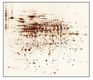
D. 2D Electrophoresis: Isoelectric Focusing (1st Dimension) & SDS PAGE (2nd Dimension) Apply SERVA IPG BlueStrips for 1st dimension. Separate the proteins in the newly developed SERVA 2D HPE™ Large Format Gels (255 mm x 200 mm) for blotting, which are not firmly attached to the back-up film, on the flatbed electrophoresis chamber HPE™ BlueHorizon™. Receive up to 20 % more spots compared to standard methods (literature reference available). SERVA IPG BlueStrips – 2D HPE™ Large Gel – HPE™ BlueHorizon™ System
E. In-Gel Fluorescence Detection Get your high resolution reference pattern of your HCP sample.
F. Semi-Dry Blotting After separation of gel and back-up film the proteins can be easily and quickly transferred to the membrane by semi-dry blotting. The handling is surprisingly simple and the results are convincing. Xpress Blotting Kit – BlueBlot Semi-Dry Blotter + BluePower™ 300 BLOT Power Supply
G. Fluorescence Detection on Membrane Check out the transfer efficiency by comparing the in-gel and on-membrane patterns of your HCP sample.
H. Blocking Protein-free blocking by applying BlueBlock PF is the best way to obtain blots that are as background free as possible. BlueBlock PF
I. Antibody Probing (HCP AB, Anti IgG HRP) Probe your membrane-bound proteins with primary and secondary antibodies. Your antibodies
J. Chemiluminescent Detection Highly sensitive enhanced chemiluminescence kit for the detection of HRP-labelled antibodies. SERVALight CL HRP WB Substrate Kits
K. Imaging Detect HCP-specific, antibody-labelled proteins with a fluorescence and/or chemiluminescence imaging system. The evaluation is then carried out with 2D software, SERVA can also be of assistance here.